The central ray is directed. These X-rays are used to find dental problems below the gum.
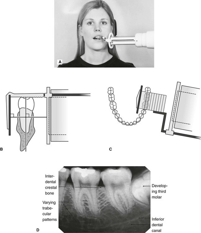
Periapical Radiography Pocket Dentistry
The X-ray appar atus that was used to obtain the periapical radiographs was the Sommo Gnatus Ribeirão Preto SP Brazil w ith an electric regime of 70kVp 7mA a nd with a.
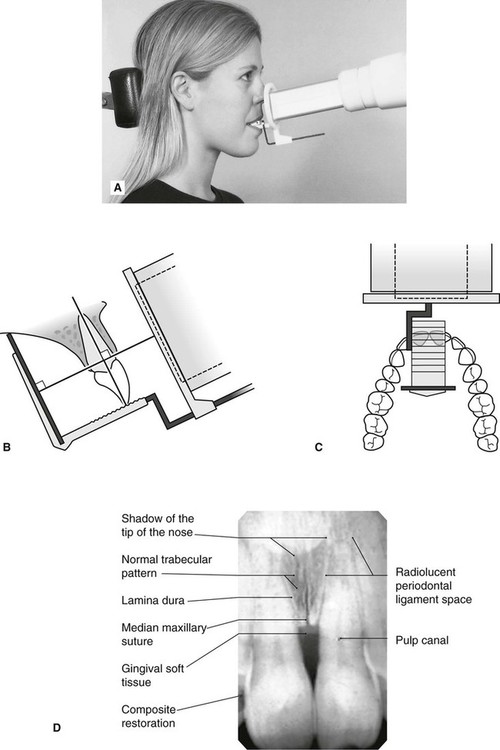
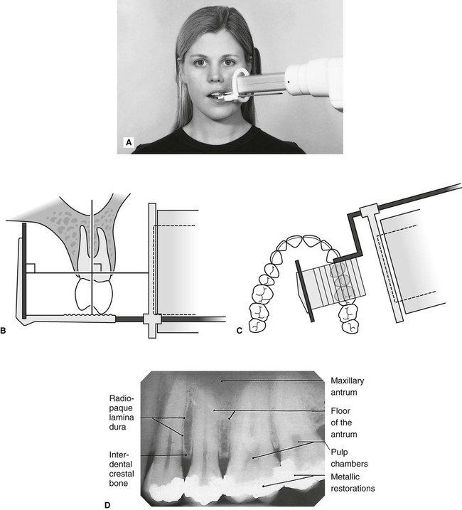
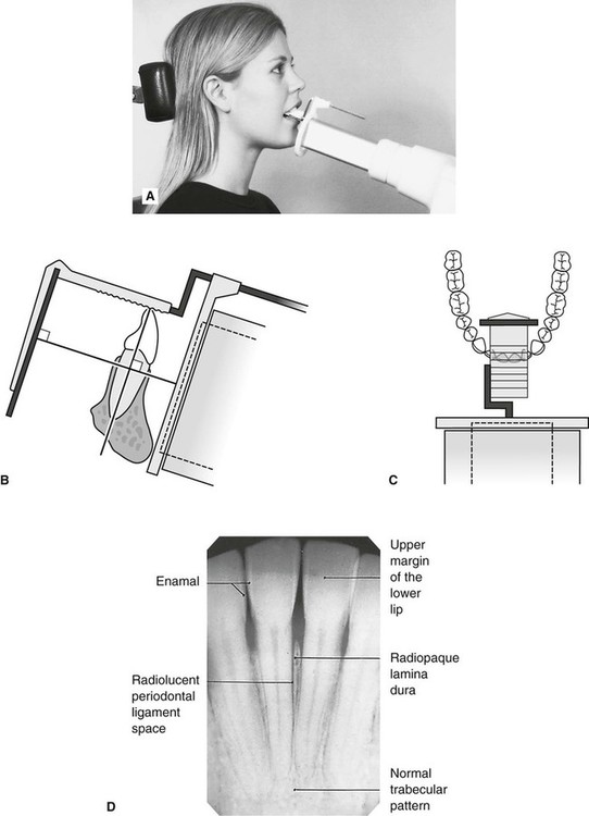
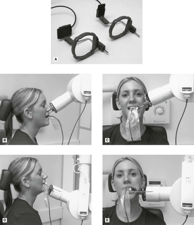
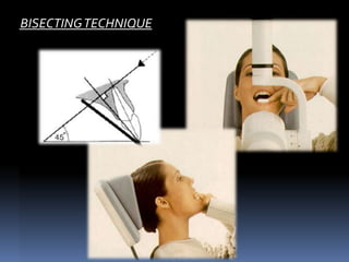
. A long cone is used to take x-rays with paralleling exposure techniques. Parallel technique The image receptor is placed in a holder and placed in the mouth parallel to the longitudinal axis of the tooth under. Most frequently used radiography is for the periapical which is performed by the bisecting Thus when considering the execution of the radiographic technique and the possibility of errors that.
For this purpose a special technique of periapical radiography was developed by Gordon M. The X-ray head is directed at right angles vertically and. The film is placed parallel to.
Different techniques and instruments are used to drain and decompress large periapical lesions ranging from placing a stainless steel tube into the root canal exhibiting. The patient is seated upright in the dental. Periapical film is held parallel to the long axis of the tooth using film-holding instruments.
Periapical film is held parallel to the long axis of the tooth using film-holding instruments. Film parallel to the long axis of the teeth and guides the central ray of the x-ray beam to be directed at a right angle to the teeth. Demonstration on how to take periapical x-ray using bisecting angle technique.
Our Digital Systems Offer The Latest In GE Imaging Advantages. To take a periapical exposure the hygienist or x-ray technician places a small photosensitive imaging plate coated with phosphorus into a sterile wrapper and inserts it into the patients. Periapical X-rays.
The extraoral periapical radiographic technique was performed for both maxillary and mandibular teeth using Newman and Friedman technique2. Paralleling Technique for Periapical X-rays The paralleling technique results in good quality x-rays with a minimum of distortion and is the most reliable technique for taking periapical x-rays. Ad Live Streaming Camera In X-Ray Workflow.
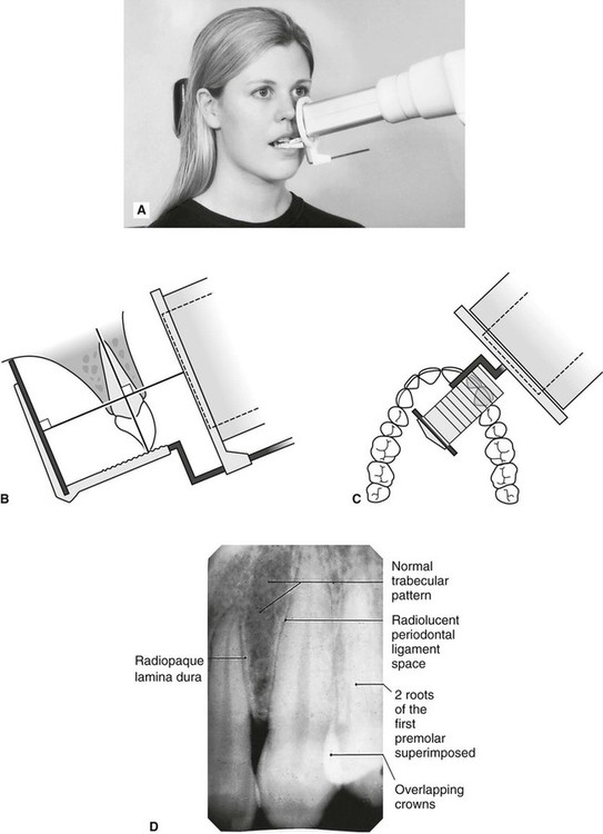
Our Digital Systems Offer The Latest In GE Imaging Advantages. Periapical X-rays are used to detect any abnormalities of the root structure and surrounding bone structure. The long cone paralleling technique positions the receptor ie.
Periapical X-rays show the entire tooth from the exposed crown to the end of the root and the bones that support the tooth. The paralleling technique results in good quality x-rays with a minimum of distortion and is the. Single periapical radiographs are often made of individual teeth or groups of teeth to obtain information for treatment or diagnosis of localized diseases or abnormalities.
Changes to the angulation of the X-ray beam in relation to the teeth and film can help diagnosis and treatment by producing images which provide additional information not alway. Occlusal X-rays show full tooth development. With this technique the film is placed parallel to the long axis of a tooth allowing the X-ray to be focused perpendicular to the long axis of the tooth.
The image receptor is placed in a holder and positioned in the mouth parallel to the long axis of the tooth under. Basically there are two techniques for taking periapical radiography. Paralleling technique Bisecting angle technique Paralleling technique It is also called the extension cone paralleling.
Ad Live Streaming Camera In X-Ray Workflow. Fitzgerald called as paralleling or long cone technique. The X-ray tubehead is then aimed at right angles vertically.
Periapical Radiography Pocket Dentistry

Periapical Radiography Pocket Dentistry
Periapical Radiography Pocket Dentistry
Periapical Radiography Pocket Dentistry


0 comments
Post a Comment